Back Bones Diagram : Skeleton Back Bones Diagram : Skeleton Anatomy System ... : Studying a spine diagram is one way to better understand many of the individual components of the back bone and how they might relate to a symptomatic back, neck or sciatica pain condition.
byAdmin•
0
Back Bones Diagram : Skeleton Back Bones Diagram : Skeleton Anatomy System ... : Studying a spine diagram is one way to better understand many of the individual components of the back bone and how they might relate to a symptomatic back, neck or sciatica pain condition.. We will attempt to provide a simplified overview of this complex anatomy. Human backbone diagram, bone, human backbone diagram. Muscle or tendon injuries can occur anywhere in the body. Stress fracture treatment symptoms u0026 causes. It contains the osteology, arthrology and myology of the spine and back.
This human anatomy module is composed of diagrams, illustrations and 3d views of the back, cervical, thoracic and lumbar spinal areas as well as the various vertebrae. Cervical spine anatomy is quite complex. The bones of the pelvis and lower back work together to support the body's weight, anchor the abdominal and hip muscles, and protect the delicate vital organs of the vertebral and abdominopelvic cavities. Bones of the neck picture. Bone diagram forehead (frontal bone) nose bones (nasals) cheek bone (zygoma) upper jaw (maxilla) lower jaw (mandible) breast bone (sternum).
Performance U. Fitness Continuing Education | An Inside ... from nicktumminello.com See lumbar spine anatomy diagram stock video clips. It also covers some common conditions and injuries that can affect the back. Spinal vertebrae bone spine vertebra toracica spinal cord spine structure back diagram spine sections spinal cord vertebrae spinal structure health diagram. Muscle or tendon injuries can occur anywhere in the body. Powerful muscles that move the head and arms attach to these bones as well. We also discuss what are osteons, what are canaliculi. Fishbone diagrams, aka ishikawa diagrams are used across various industries to analyze causes and their effect. At the back of each bone in the spine (vertebra) are bony points called processes, which muscles attach to.
Lateral labeled diagram of the human vertebral spinal column showing vertebrae numbering order and the 5 different regions of the spine.
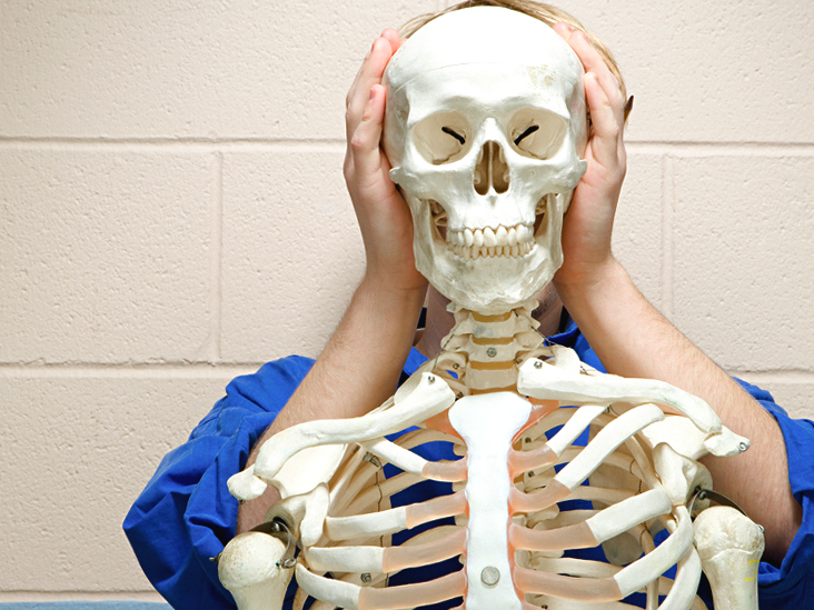
We also discuss what are osteons, what are canaliculi. Diagramme schnell und einfach erstellen. The human back extends from the buttocks to the posterior portion of the neck and shoulders. The lumbar spine connects to the thoracic spine above and the hips below. Each typical vertebra consists of a body, an arch and three processes that stem from. Daniel nelson on january 1, 2019 2 comments 🔥! The bones of the chest and upper back combine to form the strong, protective rib cage around the vital thoracic organs such as the heart and lungs. Diagram of a human female skeleton, back view. Skeleton back bones diagram / human skeleton anatomy vintage 1940s high res digital image / in this assignment, students color the various parts of the skeletal system and then answer some follow up teach your students the names of the bones in the human body with the help of this illustrated human skeleton diagram. The notochord present in the embryonic stage is replaced by the vertebral column. Fishbone diagrams, aka ishikawa diagrams are used across various industries to analyze causes and their effect. The spine or backbone consists of 26 small bones or vertebrae. It is designed to be incredibly strong, protecting the highly sensitive nerve roots, yet highly flexible, providing for mobility on many different planes.
Cervical spine anatomy is quite complex. A tough, springy disc of cartilage sits between the vertebrae of your spine. The atlas is the topmost vertebra, and along with c2, forms the joint connecting the skull and spine. The vertebral column is a part of the axial skeleton, which comprises the skull, ribs and sternum other than the vertebral column. Anatomical diagrams of the spine and back.
Human Anatomy Body - Page 3 of 160 - Human Anatomy for ... from www.anatomylibrary99.com In the back and elsewhere in the body, tendons attach muscles to bones. The red lines point individual bones and the names are writen in singular, the blue lines conect to group of bones and are in plural form. Its appearance is different from the other spinal vertebrae. Cross section of human bone diagram 12 photos of the cross section of human bone diagram cross section diagram of human bone, bone, cross section diagram of human bone. The rib cage also anchors the bones of the head, neck, shoulders, and arms to the. Human backbone diagram, bone, human backbone diagram. It contains the osteology, arthrology and myology of the spine and back. The vertebral column is a part of the axial skeleton, which comprises the skull, ribs and sternum other than the vertebral column.
Bones of the pelvis and lower back.
Cross section of human bone diagram 12 photos of the cross section of human bone diagram cross section diagram of human bone, bone, cross section diagram of human bone. These bones work together to provide. The vertebrae, which stack like spools of thread, support the back and protect the spinal cord. Bones, discs, and joints in your lower back. This vertebra supports the skull. When looking from behind, in most individuals, the spine looks straight. Lower limbs (60 bones, 30 each side). The rib cage also anchors the bones of the head, neck, shoulders, and arms to the trunk of the body. The muscles, bones, ligaments, and tendons in the back can all be injured and cause back. Lower back bones diagram : Cervical spine anatomy is quite complex. There are seven cervical vertebrae that allow for a great amount of motion in the neck. It is particularly interesting for physiotherapists.
The rib cage also anchors the bones of the head, neck, shoulders, and arms to the. Bones of the neck picture. The first seven bones (vertebrae) of your spine form your neck. Anatomical diagrams of the spine and back. The spine diagram the spine diagram shown below, consists of many bones or vertebrae,soft discs,the spinal cord, and spinal nerves.
Flat Bones: Definition, Examples, Diagram, and Structure from www.healthline.com Bones of the neck picture. The human back extends from the buttocks to the posterior portion of the neck and shoulders. The vertebrae, which stack like spools of thread, support the back and protect the spinal cord. Arms and hands bones names. Bones of the pelvis and lower back. And coccygeal the tail bone. Human backbone diagram, bone, human backbone diagram. Fishbone diagrams, aka ishikawa diagrams are used across various industries to analyze causes and their effect.
The rib cage also anchors the bones of the head, neck, shoulders, and arms to the trunk of the body.
Lateral labeled diagram of the human vertebral spinal column showing vertebrae numbering order and the 5 different regions of the spine. They help support particular bones and make them move. In the back and elsewhere in the body, tendons attach muscles to bones. It is also known as the vertebral column. Bone structure birds 12 photos of the bone structure birds bone structure birds, bone structure in. Powerful muscles that move the head and arms attach to these bones as well. Muscle or tendon injuries can occur anywhere in the body. Vertebrae are the structural constituents of the spine.there are 33 vertebrae in total; (temporal bone) shoulder blade (scapula) lower back vertebrae (5) (lumbar vertebrae) back of skull (occipital bone) fused vertebrae (5) (sacrum) hand bones (metacarpals) finger bones The first seven bones (vertebrae) of your spine form your neck. The rib cage also anchors the bones of the head, neck, shoulders, and arms to the trunk of the body. Related posts of human back bones diagram bone structure birds. It is designed to be incredibly strong, protecting the highly sensitive nerve roots, yet highly flexible, providing for mobility on many different planes.
The rib cage also anchors the bones of the head, neck, shoulders, and arms to the trunk of the body back bones. The bones of the pelvis and lower back work together to support the body's weight, anchor the abdominal and hip muscles, and protect the delicate vital organs of the vertebral and abdominopelvic cavities.